PHONE
+44-7482-878921
+44-7482-878921
2376-0249
Case Blog - International Journal of Clinical & Medical Images (2016) Volume 3, Issue 4

Author(s): N Selvakumar, Neerav Goyal, Sandeep Vohra and Subash Gupta
Abstract: Hepatic arterial anomalies are well studied and classified. Still we come across surprises in liver surgeries. Thanks to the imaging modalities that these anomalies are no more a surgical surprise. We describe one such case of hepatic arterial anomaly encountered while a right donor hepatectomy. The right hepatic artery instead of entering the porta through the hilar plate was entering through the cystic plate and then running a short subcapsular course joins the portal pedicle. The artery could have been easily damaged as a cystic artery or hilar plate artery had it not been properly studied preoperatively. There was no difficulty in the implantation. Post operatively there were no complications in general and arterial complications to be specific.
Introduction: The raising number of living donor liver transplantations has improved the understanding of vascular and biliary anomalies in liver. There are standard anatomical and surgical classifications for hepatic artery, portal vein and bile duct [1,2]. Still unclassified variants are often encountered. In donor hepatectomy where the margin of error is very very less these unclassified variants are of great significance. We describe one such hepatic arterial anomaly that we encountered recently out of over 1900 LDLTs done in our institute so far. Case presentation Our case was a 21 years old healthy male from Pakistan. He was accepted as a healthy liver donor for his 48 years old uncle who had HCV related decompensated CLD.
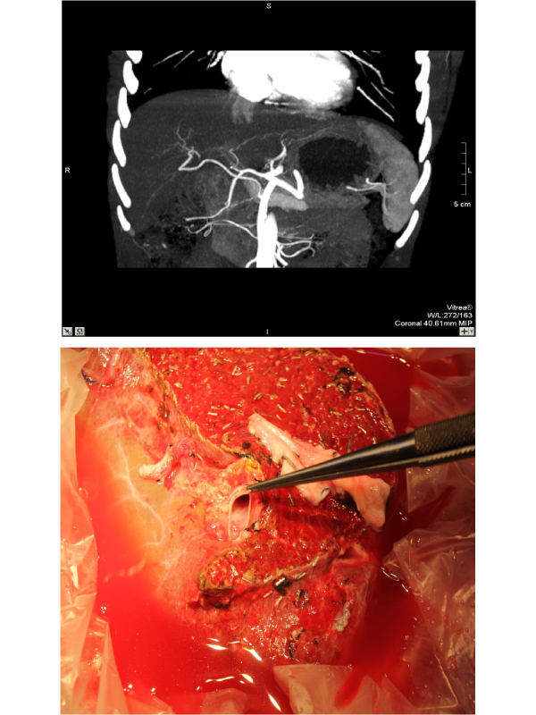
The donor on further evaluation with CTLA had healthy non fatty liver with adequate remnant with replaced RHA, type 1 portal vein and Type a biliary anatomy. The hepatic artery in addition to being replaced had an unusual hump at the level of entry into the liver (Figure 1). Instead of entering the liver at the hilar plate the artery was entering the liver at the cystic plate.
The segment 6 artery was seen arising from the entry point. Later the main artery made a medial turn before making the portal triad. This anomaly was well appreciated and studied and the surgery performed carefully to avoid any damage. The graft liver with the anomalous right hepatic artery is depicted in Figure 2. Implantation was done with anastomosis to the recipient right hepatic artery. Post operatively the graft function was good. The patient had uneventful recovery.
 Awards Nomination