Introduction: Patients presenting to the emergency room with colorectal foreign bodies are uncommon, but not rare events. The majority of published reports about rectal foreign bodies are case reports and case series. Foreign bodies in the colorectal region can cause a variety of complications, including death. Because patients are embarrassed, they often attempt to remove the foreign body at home and delay seeking medical treatment.
Case presentation: We present a case of a 60-year-old white male with a colorectal foreign body for 5 days, that he was unable to remove after multiple maneuvers at home. After multiple unsuccessful attempts, operative intervention with laparotomy and colostomy with removal of the foreign body was done.
Conclusion: Our case represents a 24 cm foreign body colorectal insertion presenting to emergency room after multiple failed removal attempts at home. The foreign body was deep into the sigmoid colon. This case highlights the importance of evaluating the removal approach based on the assessment of edema sounding the foreign body.
Introduction: Reports of people inserting foreign bodies into the rectum, for a variety of reasons, have appeared as early as the 16th century [1]. There has been increase in the incidence of these types of events reporting to emergency rooms in recent years. There are a number of complications that can arise from foreign bodies in the colorectal region, including pain, constipation, bowel perforation, sepsis, and death. Delay in seeking care commonly happens because the patient attempts to remove the object at home due to the associated embarrassment. Here we present a case of colorectal foreign body.
Case Report: A 60-year-old male was seen in emergency room with the chief complaint of having a glass Coke bottle lodged in his rectum, with the bottle top facing up. His girlfriend had introduced this voluntarily for sexual purposes 5 days prior. Since that time, he had been having intermittent, ongoing, crampy abdominal pain. He had tried laxatives, excessive food and fluid intake, along with attempts at manual removal, all with no success. He did not have fever, nausea, vomiting, or rectal bleeding.
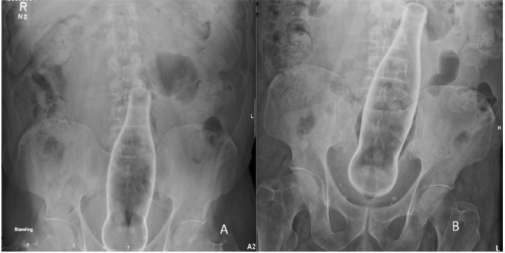
On presentation, the abdomen was soft, and there was no evidence of sepsis. Abdominal radiograph showed a sharply delineated glass bottle, 24 cm x 5.5 cm, positioned over the lower abdomen and pelvis (Figures 1A and 1B). No free air was noted. The rectal examination showed the bottom of bottle approximately 7 cm from the anal verge with no lacerations or sphincter. Transanal removal was attempted under general anesthesia in the operating room. There was swollen prolapsing tissue with edema noticed on examination with retractors. Multiple attempts at transanal removal failed. Gentle lower abdominal pressure helped move the foreign body down to about the pelvic inlet musculature, but it became tightly wedged at that level. A Foley catheter was inserted, and air was infused in an attempt to break any vacuum pockets behind the foreign body. The balloon was then inflated in an attempt to pull the foreign body down. This was unsuccessful. Further attempts to secure the bottle with large, rubber-shod clamps were also unsuccessful, as the foreign body would not engage. Obstetrical vacuum forceps and obstetrical forceps were tried, which also could not bring the foreign body down.
After multiple unsuccessful attempts, it was recognized the edema was making it difficult to remove the foreign body using the transanal approach. Therefore, operative intervention with laparotomy and colostomy with removal of the foreign body was done. The sigmoid colon was inflamed and erythematous, but no perforations were evident. Segmental sigmoid resection and Hartmann’s procedure were undertaken given the contamination along with the inflammation, which was evident preventing it from primary anastomosis.
The patient’s hospital course was complicated by an abdominal wound infection with dehiscence and deep venous thrombosis of his lower extremities. He was treated with wound re-exploration and re-closure of the wound dehiscence with retention sutures along with negative pressure wound therapy and with anticoagulation therapy for the deep venous thrombosis. Subsequently, the patient recuperated well in rehabilitation and was discharged to home.
Discussion: People place foreign bodies in the rectum for various reasons: sexual vs. nonsexual, voluntary vs. non-voluntary. The vast majority is inserted for autoerotic purposes, but it may also be due to assault or trauma, or could be placed iatrogenically such as thermometers or enema tips. Additionally, in drug trafficking, illicit drugs (cocaine, amphetamine and marijuana) are packed in small plastic bags and placed in the rectum, which can be encountered in an emergency room setting. Uncommonly, objects could be lodged in the colon from perioral ingestion. Various retained objects recorded in the literature include sex toys, tools and instruments, bottles, cans, jars, poles, pipes and tubing, fruits and vegetables, stones, balls, balloons, umbrellas, light bulbs, and flashlights [2]. There have been case reports of retained rectal foreign bodies of up to 5 years [3].
Classification of the level of entrapment has helped stratify the likelihood of transanal extraction. Those in the low or mid rectum, up to a level of 10 cm, most often can be removed transanally. Those above this level, in the upper rectum, may require laparotomy for retrieval [4]. It has been demonstrated that objects located in the sigmoid colon are 2.5 times more likely to require operative intervention versus those located more distally in the rectum [5]. Another classification system stratifies into four categories based on the extent of injury. Category 1–retained foreign body without injury, and Category 2-non-perforative mucosal laceration, are the most straight forward. Sphincter injury and recto-sigmoid perforation represent Categories 3 and 4, denoting much more serious injuries [2,6]. Obtaining a complete and thorough history is necessary, as the patient may be likely to fabricate a story, concealing the true story due to embarrassment. Sometimes clues for involuntary placement of objects should be sought to verify an abuse/assault situation. A high degree of suspicion should be considered for retained medical instruments, especially in children and the mentally ill. The first thing to look for in a physical examination is any sign of peritonitis and to determine whether the patient is unstable. If so, they warrant an emergent laparotomy, and no attempts at bedside removal should be undertaken [7]. Resuscitation with intravenous fluids and antibiotics may be needed depending upon the individual presentation. A rectal examination also provides valuable information on the location of the object and aids in assessment of any sphincter damage. Acute sphincter damage was shown to have good long-term prognosis in one small study [4]. Abdominal radiography might give clues on the location and shape of the object to assist in removal and also to rule out free air from full thickness injury.
There are methods of extraction: transanal, endoscopic, and operative. Some suggest waiting for the object to pass spontaneously [8], however, this usually does not occur, and attempts at removal should be undertaken regardless of the object’s location [7]. The transanal approach is successful in 60%–75% of cases [6-9]. Use of a perianal block and conscious sedation helps in better relaxation of the anal sphincter, increasing the chances of successful removal. Additionally, suprapubic pressure applied by an assistant helps in pushing the object caudally. In awake patients, having them perform the Valsalva maneuver works in the same manner. Various tools such as ring forceps, obstetric forceps, and suction devices have been used. A vacuum is created above the object, which sucks the object in, and prevents it from coming down. To overcome this, a balloon catheter (such as a Foley) can be inserted beyond the object, creating an infusion of air that helps break the vacuum, allowing easier retrieval of the object [7]. The inflated balloon can also be used to pull the object down. It is cautioned to avoid using graspers in body drug packers to prevent breakage of plastic bag containing the drugs; in these circumstances, only digital removal should be attempted. Other documented attempts at retrieval include using plaster of Paris or super glue to hold objects and also drilling holes in the objects [6-8]. Caution should be taken with maneuvers of glass foreign bodies to avoid breaking the glass. If the above-described attempts are unsuccessful, endoscopy can be used. Polypectomy snare, biopsy forceps, guide wire, and balloon techniques can also be used [10]. These may require using fluoroscopy techniques [11]. Endoscopic techniques are helpful for smaller foreign bodies. If all of the above attempts are unsuccessful, general anesthesia can be utilized. Attempts at using transanal and endoscopic methods will have better anal sphincter relaxation with general anesthesia. If unsuccessful, colostomy can be done. Hartman’s procedure should be performed if there is gross contamination from the object; otherwise primary anastomosis could be attempted if there is good tissue quality [12].
Post-extraction management after successful removal includes rigid proctoscopy and/or flexible sigmoidoscopy to identify any mucosal damage, active bleeding, bowel injury, or additional retained objects. Abdominal radiography should be performed after removal of the object to rule out perforation. There have been no long-term studies done on colorectal foreign bodies. One case series followed 30 patients after colorectal foreign body removal, and none reported long-term incontinence to solid, liquid, or gas over a time period ranging from 8 to 96 days [13]. Sphincter dysfunction that may be present after foreign body removal usually improves with observation; however, if incontinence persists, delayed sphincteroplasty may be performed [4].
Conclusion: Our case represents a 24 cm glass foreign body that was inserted colorectally in a patient who presented to the emergency room after 5 days. He had used the object for voluntary autoerotic purposes and was forthcoming with the history. He had failed multiple attempts at home to remove the object. The foreign body was deep into the sigmoid colon and was classified as a Category 1 without lacerations or perforations at presentation, although significant edema surrounding the object prevented transanal removal. An endoscopic approach was not attempted as the object was too large for an endoscopy snare or forceps and also given the edema sounding the foreign body. It was necessary to take caution to not break the glass. The patient subsequently had a sigmoid colectomy and Hartman’s procedure because of gross contamination.


 Awards Nomination