2376-0249
Clinical-Medical Image - International Journal of Clinical & Medical Images (2023) Volume 10, Issue 5

Author(s): Merbouh Sahar*, Kaoutar Imrani, Amine Cherraq, Nabil Moatassimbillah and Ittimade Nassar
Department of Radiology, CHU IBN SINA University Hospital, Mohammed V University, Rabat, Morocco
Received: 13 April 2023, Manuscript No. ijcmi-23-95512; Editor assigned: 14 April 2023, Pre QC No. P-95512; Reviewed: 28 April 2023, QC No. Q-95512; Revised: 03 May 2023, Manuscript No. R-95512; Published: 10 May 2023, DOI:10.4172/2376-0249.1000894
Citation: Sahar M, Imrani K, Cherraq A, Moatassimbillah N and Nassar I. (2023) Retroperitoneal Fibrosis. Int J Clin Med Imaging 10: 894.
Copyright: © 2023 Sahar M, et al. This is an open-access article distributed under the terms of the Creative Commons Attribution License, which permits unrestricted use, distribution, and reproduction in any medium, provided the original author and source are credited.
Retroperitoneal fibrosis (RPF) is a rare disease characterized by the presence of aberrant fibroinflammatory tissue forming in the retroperitoneal space surrounding the subrenal aorta, the iliac arteries and the adjacent structures. It is most often idiopathic, sometimes it can be secondary to infections, abdominal surgery, medication or retroperitoneal tumors (primary or metastatic). Imaging plays an important role in the diagnosis of retroperitoneal fibrosis, it allows to distinguish between benign and malignant forms, and also to follow the evolution under treatment.
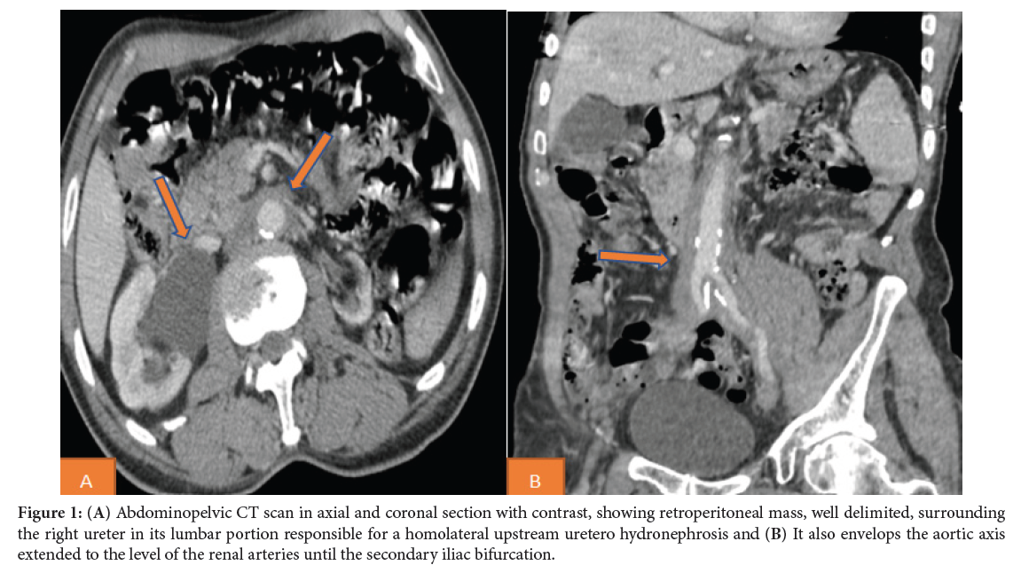
Retroperitoneal fibrosis (RPF) is a rare disease characterized by the presence of fibro-inflammatory tissue surrounding the retroperitoneal structures. It is idiopathic in the majority of cases [1]. Its etiopathogeny remains obscure. According to current data, the etiopathogeny of RPF is part of a fibro-inflammatory systemic disease called “IgG4 disease” with involvement of different organs [2]. Clinical signs are highly variable and non-specific, often related to the mechanical effect of the RPF on adjacent structures. The diagnosis of retroperitoneal fibrosis is made on the basis of imaging data and biological arguments. Biological exploration classically finds an elevation of inflammation markers associated with an inflammatory anemia and an alteration of the renal function. Anti-nuclear antibodies (ANA) and other autoantibodies may be positive [3] Serum IgG4 may be elevated in patients with retroperitoneal fibrosis related to IgG4 disease [4]. CT and MRI are the examinations of choice for the diagnosis and distinction between benign and malignant forms. They reveal the fibrosis plaque, specify its morphology, its location and its propagation to the adjacent structures. The initial fibrosis begins near the aorta and iliac arteries, extends through the retroperitoneum to involve the ureters. The center of fibrosis is usually located at the aortic bifurcation; this abnormal tissue bifurcates to follow the common iliac arteries [5]. On the CT scan, retroperitoneal fibrosis is most often manifested by a paraspinal retroperitoneal mass, well delimited, irregular, that is isodense to surrounding muscle. The degree of soft-tissue enhancement on CT correlates with activity of the fibrotic process. MRI shows, at the early, inflammatory stage a T2 hypersignal and an intense enhancement related to hypercellularity-edema, the late, inactive stage shows a weak T2 signal and weak or absent enhancement. The PET is a well-established functional imaging modality in oncology and is increasingly used in the evaluation of inflammatory diseases, including RPF. It has a very high sensitivity superior to CT or MRI. It allows a comprehensive assessment of the inflammatory involvement in idiopathic form, assesses the intensity of the inflammatory process in active phase, allows assessment of response to treatment and can predict the prognosis. Biopsy is not always performed, especially if the radiological features are consistent with RPF. Biopsy is only indicated in atypical forms or unusual locations. When considered, surgical biopsy is preferred to obtain a larger quantity of material for a more reliable histological analysis. Treatment is based on corticosteroids and surgery. In case of dilatation of the urinary excretory tract, disobtruction by double J catheter or percutaneous nephrostomy is necessary during the acute phase in association with medical treatment. The prognosis of the disease is favorable, but the risk of recurrence requires prolonged clinicobiological and radiological surveillance [1-5].
Retroperitoneal fibrosis; Diagnosis; Imaging
The authors declare that they have no conflict of interest in relation to this article.
[1] Saihi M, Chargui S, Hamida FB and Abdallah TB. (2018). La fibrose rétropéritonéale. Nephrol Ther 14: 393.
Google Scholar, Crossref, Indexed at
[2] Tang CSW, Sivarasan N and Griffin N. (2018). Abdominal manifestations of IgG4-related disease: A pictorial review. Insights Imaging 9: 437-448.
Google Scholar, Crossref, Indexed at
[3] Kermani TA, Crowson CS, Achenbach SJ and Luthra HS. (2011). Idiopathic retroperitoneal fibrosis: A retrospective review of clinical presentation, treatment and outcomes. Mayo Clin Proc 86: 297-303.
Google Scholar, Crossref, Indexed at
[4] Zen Y, Onodera M, Inoue D, Kitao A and Matsui O, et al. (2009). Retroperitoneal fibrosis: A clinicopathologic study with respect to immunoglobulin G4. Am J Surg Pathol 33: 1833-1839.
Google Scholar, Crossref, Indexed at
[5] Cronin CG, Lohan DG, Blake MA, Roche C and McCarthy P, et al. (2008). Retroperitoneal fibrosis: A review of clinical features and imaging findings. AJR Am J Roentgenol 191: 423-431.
 Awards Nomination

