PHONE
+44-7482-878921
+44-7482-878921
2376-0249
Case Blog - International Journal of Clinical & Medical Images (2015) Volume 2, Issue 12

Author(s): Animesh Mishra, Tony Ete*, Rinchin Dorjee, Pravin Jha, Gaurav Kavi, Amit Malviya
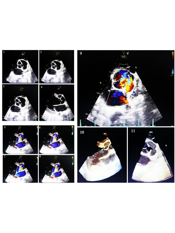
Case Presentation: 29 years old male got admitted following complaints of palpitation and coughing for one year. Patient also had complaints of difficulty in breathing for one year that was gradual in onset and intermittent chest pain for the same duration. He also had history of paroxysmal nocturnal dyspnoea (PND) in between for one year. On examination, pulse rate was 96/minute, regular, high volume, BP-140/60 mmHg. Cardiovascular system examination revealed shifting of apex laterally in the fifth left intercostals space. Patient had continous murmur on ausculatation in the second and third left intercostal space. Rest of the system on examination revealed no significant abnormality. Routine investigations revealed Hb-12.9 gm%, TLC-7800/cumm, platelets-2.7 lakhs/cumm, Urea-26 mg/dl, Creatinine-0.6 mg/dl, normal liver function test. Blood for HbsAg, Anti HCV and HIV 1/HIV 2 antibody were negative. Chest X ray showed Cardiomegaly. Echocardiography revealed aneurysm of left coronary sinus withruptured left sinus of valsalva draining into left ventricle. Left atrium, Left ventricle and right atrium were dilated. It also showed severe mitral regurgitation, mild tricuspid regurgitation and pulmonary artery systolic pressure of 75 mmHg. Left ventricular ejection fraction was 40%. No intracardiac clot, vegetations or pericardial effusion was present. Initially patient was managed conservatively with diuretics and beta blockers. Once the patient was stabilized patient was sent to cardiothoracic vascular surgery department for surgical correction (Figures 1-3).
Discussion: Ruptured sinus of Valsalva (RSOV) aneurysms are rare comprising 0.3-3.56 % of all congenital heart diseases [1]. The right sinus of Valsalva is most commonly involved and usually ruptures into right heart chambers. Involvement of left heart chambers is very uncommon. Rupture of left sinus of valsalva is very uncommon. Apart from that rupture of LSOV into left ventricle is a very rare finding [2]. Aneurysms of sinus of Valsalva are usually thought to result from absence of normal elastic and muscular tissue in the aortic sinus leading to thinning of its wall [3]. Presentation of sinus of Valsalva aneurysm is usually varied. Patient may present with chest pain and dyspnea. Chest pain is due to acute AR from the ruptured sinus of Valsalva, frank or dynamic coronary artery compression, or dissection into a coronary artery.
Unruptured sinus of Valsalva aneurysms is usually asymptomatic. However it may present with right ventricular outflow tract obstruction when they bulge into right ventricular outflow tract. It may also present with complete heart block if it erodes into interventricular septum. Diagnosis of ruptured sinus of valsalva is usually done with noninvasive methods like echocardiography, computerized tomography and magnetic resonance imaging. Treatment is usually done with repair of the aneurysm with replacement of the aortic valve [4].
 Awards Nomination