PHONE
+44-7482-878921
+44-7482-878921
2376-0249
Clinical Image - International Journal of Clinical & Medical Images (2018) Volume 5, Issue 4

Author(s): Youness Jabbour*, Amine Slaoui, Tarik Karmouni, Khalid El Khader, Abdellatif Koutani and Ahmed Iben Attya Andaloussi
Abstract: Scrotal calculi, also known as scrotal pearls, are benign incidental extra testicular macro-calcifications within the scrotum. Hydrocele and repeated micro-trauma may be risk factors for development of scrotal calculi. They are usually of no clinical significance but literature data suggest their role in scrotal pain. Keywords: Scrotal calculi; Ultrasonography; Hydrocele Introduction Scrotal calculi correspond to the presence of freely floating or located calcifications lying between the layers of the tunica vaginalis of the testes [1]. It’s a rare discovery with a reported incidence ranging between 1.5% and 3% [1,2]. Intrascrotal calculi was first found by Kickham during surgery in 1935 and he described it as a “fibrinoid loose body” or “scrotal pearl”
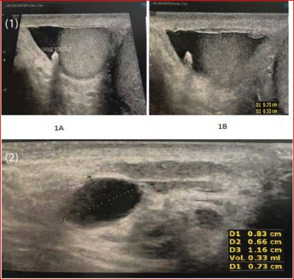
Case Presentation: We report the case of a 38 years old patient with no specific medical history complaining about an intermittent scrotal pain. Clinical examination found a swelling of the right spermatic cord without cough impulse. No other abnormalities of genitalia were noticed. Scrotal ultrasonography found both testis of normal size with the presence of 0.73 × 0.32 cm mobile scrotal calculi in the right hemiscrotum associated a small hydrocele (Figure 1A and 1B). A right epidydimal cyst of 0.83 × 0.66 × 1.16 cm was also found (Figure 2). The patient received analgesic treatment with good evolution and was kept under close follow-up.
Discussion: Scrotal calculi are mostly incidental finding in patients during scrotal ultrasound examination and easily recognized given the hyper echoic nature of the calcification which causes a discrete acoustic shadow. Detection rate of scrotal calculi is gradually increasing since they are well visualized with the application of high-frequency ultrasound. They can also be detected during surgery, on self-palpation of the testis by patients. The etiology of scrotal calculi is unclear. They may develop as a sequela to trauma or inflammatory diseases affecting the scrotum or from detached portions of the appendices testis or epididymis after torsion or infarction [1,4]. Literature data reports a high association between hydrocele and scrotal calculi suggesting its role in the genesis of scrotal calculi by crystallization of contains of the scrotal fluid. Tan et al. reported an association of hydrocele and scrotal calculi exceeding. Hydrocele was the most common additional scrotal abnormality (29.8%) in Aslan et al. study.
Frauscher et al. reported scrotal calculi as the most common diagnosed scrotal abnormality with a high incidence of 81% suggesting the role of repeated chronic micro-trauma of the scrotal contents in the genesis of scrotal calculi. Patients with an association of hydrocele and scrotal calculi exceeded 50% in Tan et al. study. Hydrocele was the most common additional scrotal abnormality (29.8%) in Aslan et al. study. Clinical significance of scrotal calculi is uncertain but some authors suggested their participation in scrotal pain given the presence of scrotal pain in patients with scrotal calculi who had no know additional scrotal abnormalities [2-5]. This pain has been explained by the movement of calculi in the fluid between the layers of the tunica vaginalis which may irritate the surrounding soft tissue, resulting in occasional pain. To date, no information is available in the literature about the results of chronic irritation due to scrotal calculi.
 Awards Nomination