PHONE
+44-7482-878921
+44-7482-878921
2376-0249
Clinical-Medical Image - International Journal of Clinical & Medical Images (2022) Volume 9, Issue 1

Author(s): Hatim Essaber*, Meryem Echchikhi, Moatassim billah Nabil, Ittimade Nassar
Received: 07 January, 2022, Manuscript No. ijcmi-22-53962; Editor assigned: 08 January, 2022, PreQC No. P-53962; Reviewed: 13 January, 2022, QC No. Q-53962; Revised: 18 January, 2022, Manuscript No. R-53962; Published: 23 January, 2022, DOI: 10.4172/2376-0249.1000806
Clinical-Medical Image
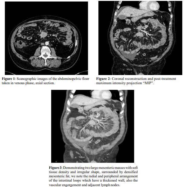
Clinical-Medical Image The mesenteric carcinoid tumor is a rare neuroendocrine tumor that can involve all organs, but which preferentially sits in the gastro-mesenteric tract with a frequency of 60 to 70%, and a predilection for the small intestine. On CT, the diagnosis of mesenteric carcinoid tumor is based on a typical triad associating an often calcified mesenteric mass; a parietal thickening of the small intestine in contact with this mass and the infiltration of the adjacent fat “in the radius of a wheel” (Figures 1-3). The wheel spoke appearance corresponds to linear thickening of the mesenteric fat diverging from the tumor mass and producing a radial appearance at the periphery. This aspect results from a microscopic tumoral extension within the mesentery, responsible for a perivascular inflammatory phenomenon then fibrotic at the level of the mesenteric fat. These results in vascular engorgement and radial densification of the mesenteric fat converging on the tumor mass and giving the typical stellar appearance at the level of the mesentery, the degree of which is proportional to the amount of peri-tumoral fibrosis developed. The “wheel spoke” sign can be encountered in other pathological entities such as idiopathic mesenteric fibrosis, peritoneal carcinomatosis and lymphoma. However, its association with calcified tumor mass and thickening of the intestinal wall is specific to carcinoid tumors
Keywords: Vertebral haemangioma; Polka-dot sign; Corduroy sign
Declaration of Conflicting Interests
The authors declared no potential conflicts of interest with respect to the research, authorship, and/or publication of this article.
References
[1] Tokpo AJ, Mohammadine B, Stitou K, Lakhdar F, Benzagmout M, et al. (2021) Unruptured Delivery of Posterior Fossa Hydatid Cyst in Child. Int J Clin Med Imaging 8:756
 Awards Nomination