PHONE
+44-7482-878921
+44-7482-878921
2376-0249
Case Blog - International Journal of Clinical & Medical Images (2016) Volume 3, Issue 8

Author(s): Aditi S Pandit and Senthilkumar Thiyagarajan
Clinical Presentation: A 72 year female presented with complaint of pain in back since 45 days radiating to both lower limb (anterior aspect) aggravated since 7 days. She went to orthopedician where she was advised for X-ray investigation and diagnosed as spondylolithesis and was referred to physiotherapy department for further treatment. She came to The Oxford College of physiotherapy outpatient department on 7th June 2016. Similar episode was experienced 15 years ago, for which she took physiotherapy treatment at Indore for 2 months and followed by one of physiotherapy treatment she felt better. So stopped to follow-up and routine exercise advised by the therapist. She is a known case of diabetes and hypertension since 35 years. She had undergone hysterectomy 30 years ago. Presently she has complaints of dull aching, intermittent pain which is aggravated on walking, standing and relieves on rest and medication. Pain on Visual Analog Scale was 8 on 10. On observation, step sign present presence of Trendelenburg gait. On palpation grade 1 tenderness over S1 level, paraspinal muscle spasm present, trigger points were present over piriformis, hamstrings, gastrocnemius.
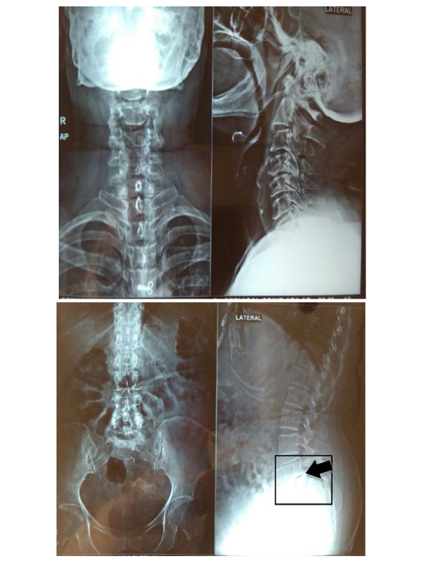
On assessment straight leg raise and prone knee bending was positive suggesting of neural tissue involvement. On X-ray investigation, osteophytes formation in cervical, thoracic and lumbar region, anterior slipping of L5 over S1 indicating spondylolisthesis. A clinical finding reveals (Figures 1 and 2) that spondylolisthesis grade III which means 50% to 75% anterior slipping of L5 vertebrae over S1. (Figure 2 with down arrow). Regular physiotherapy follow-up was advised to the patient. Physiotherapy management as follows spinal flexion exercises, core strengthening, stretching the hamstring and calf muscles. Spinal extension exercise strictly contraindicated. After the few sessions of physiotherapy, she felt comfortable.
 Awards Nomination