PHONE
+44-7482-878921
+44-7482-878921
2376-0249
Case Blog - International Journal of Clinical & Medical Images (2015) Volume 2, Issue 4

Author(s): Alex H Wong, Alexander KC Leung* and Benjamin Barankin
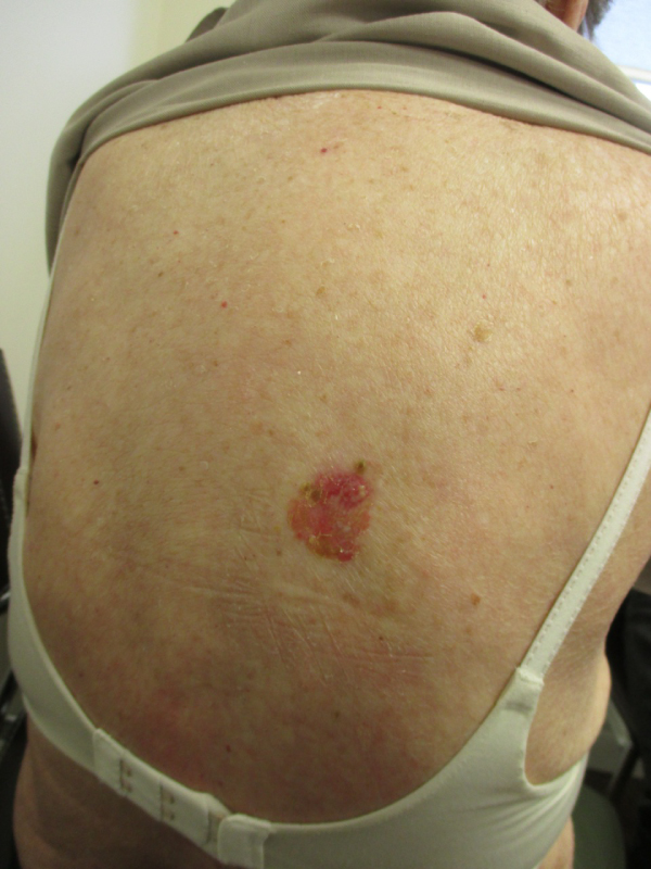
A 62-year-old Caucasian woman presented with a slowly growing, asymptomatic, red plaque on her back that was first noted a year ago. Her family physician treated her with a topical antifungal and subsequently a topical steroid without success. Basal cell carcinoma (BCC) is generally defined as a locally invasive, slowly spreading tumor that rarely metastasizes, arising in the epidermis or hair follicles and in which, in particular, the peripheral cells usually simulate the basal cells of the epidermis. BCCs are a heterogeneous group of tumors that based on their clinical morphology can be classified most commonly into the following subtypes: nodular BCC, superficial BCC, sclerosing BCC, infiltrative BCC, micronodular BCC, and pigmented BCC. Superficial BCC is the second most common subtype, accounting for 9 to 17.5% of all cases. Unlike other BCC subtypes, it tends to have an earlier age of onset and a female predominance. It is the least aggressive subtype. Typically, the lesion presents as a solitary, well-defined, slowly growing, erythematous or pinkish, scaly patch or plaque that has a thin pearly border. The size varies from a few millimeters to several centimeters in diameter. The condition is usually asymptomatic. Occasionally, the lesion may be pigmented or ulcerated. Sites of predilection include the trunk, shoulders, and extremities. Diagnosis is often delayed as it is often mistaken for a dermatitis or tinea infection. For lesions occurring at a low-risk anatomic site (e.g., trunk and extremities) with distinct clinical borders and sizes less than 2 centimeters in diameter, treatment options include surgical excision, electrodessication with curettage, liquid nitrogen cryotherapy, topical immunotherapy (e.g. imiquimod), topical 5-fluorouracil, and photodynamic therapy. Patients should have regular follow-up at least yearly following diagnosis to monitor for local recurrence or the development of new BCCs or other skin cancers at other sites.
http://www.journalinsight.org/
http://www.escientificjournals.com/
http://www.escienceopen.com/
http://www.esciencejournals.org/
http://www.esciencejournals.com/
http://www.emedicalscience.com/
http://www.emedicalsci.org/
http://www.emedicalsci.com/
http://www.microbiologyres.com/
http://www.microbialjournals.com/
http://www.immunologyres.com/
http://www.immunologyinsights.com/
http://www.molecularbiol.com/
http://www.esciencejournal.org/
http://www.oajournal.org/
http://www.journalsres.org/
http://www.journalsres.com/
http://www.journalsoa.org/
http://www.journalsoa.com/
http://www.journalsci.org/
http://www.journalres.org/
http://www.journalres.com/
http://www.journaloa.org/
http://www.journalinsights.org/
http://www.jpeerreview.org/
http://www.imedresearch.com/
http://www.imedpubjournals.com/
http://www.imedpubjournal.org/
http://www.imedjournals.org/
http://www.peerreviewedjournal.org/
http://www.peerjournals.org/
http://www.peerjournals.com/
http://www.sciencesinsight.org/
http://www.scholarresearch.com/
http://www.scholarres.org/
http://www.nutritionres.com/
http://www.gastroinsights.org/
http://www.pathologyinsights.org/
http://www.echemistry.org/
http://www.echemcentral.com/
http://www.chemistryres.com/
http://www.biochemresearch.org/
http://www.biochemjournals.com/
http://www.ebusinessjournals.org/
http://www.businessjournals.org/
http://www.peerjournal.org/
http://www.oajournalres.com/
http://www.alliedres.org/
http://www.alliedjournals.org/
http://www.alliedjournal.org/
http://www.scientificres.org/
http://www.scientificres.com/
 Awards Nomination