PHONE
+44-7482-878921
+44-7482-878921
2376-0249
Case Blog - International Journal of Clinical & Medical Images (2015) Volume 2, Issue 10

Author(s): Chi-Chou Tseng, Kuen-Huang Chen, Chien-Hung Lin and Wei-Ting Lin*
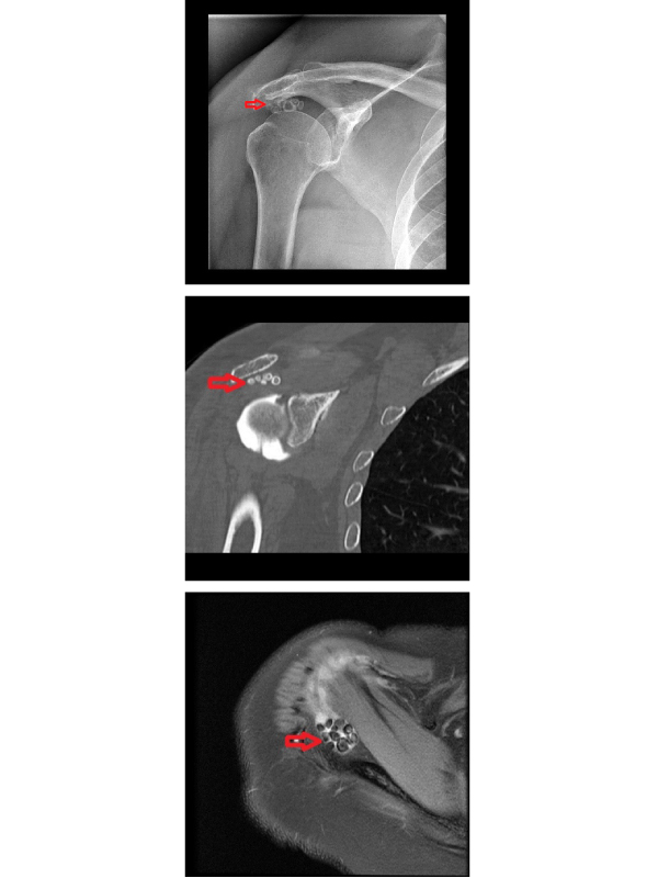
Clinical Presentation: A 54-year-old female presented with intermittent pain over right shoulder off and on for 1 month. She did not recall any associated major trauma, but she was a retired teacher who frequently needed raising right shoulder for writing on blackboard. Physical examinations only showed a swelling mass about 2 × 1 cm with mild tenderness over right shoulder joint and mild limited of range of motion. The radiography of right shoulder plain film showed multiple rounded, calcified bodies around the shoulder joint (Figure 1).
Computed tomography (CT) (Figure 2) and magnetic resonance (MR) images confirmed the presence of widespread distribution of the calcified bodies throughout the shoulder joints with partial thickness tear of supraspinatus tendon involving bursal side and intrasubstance (Figure 3). Therefore, the diagnosis of synovial osteochondromatosis was made. She only received rehabilitations for right shoulder for symptomatic treatment, and the condition gradually improved. Synovial osteochondromatosis is caused by the formation of multiple nodules of hyaline cartilage within the sub-synovial connective tissue. Most of them develop in middle age men, and knee is the most common site of involvement [1].
In addition, hip, elbow, ankle and shoulder have been reported to be involved [1]. The clinical manifestations of osteochondromatosis include painful swelling, limited range of motion, and locking, crepitation, and palpable loose bodies [2]. Surgical excision of the synovium and removal of the loose bodies for symptomatic patients remains the treatment of choice, and the prognosis is always fair in spite of rare recurrence.:
 Awards Nomination