PHONE
+44-7482-878921
+44-7482-878921
2376-0249
Clinical-Medical Image - International Journal of Clinical & Medical Images (2022) Volume 9, Issue 4

Author(s): Khairiyah Sidek*
Received: 21 April, 2022, Manuscript No. ijcmi-22-63675; Editor assigned: 23 April, 2022, 2022, PreQC No. P-63675; Reviewed: 25 April, 2022, QC No. Q-63675; Revised: 27 April, 2022, Manuscript No. R-63675; Published: 30 April, 2022, DOI: 10.4172/2376-0249.1000824
Clinical-Medical Image
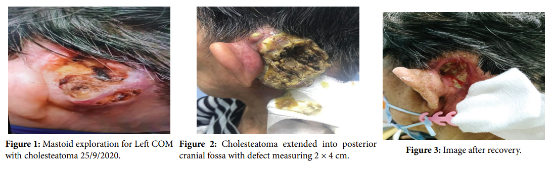
77 years Chinese female, underlying hypertension, history of left ear surgery at age 10 years old presented with left sided facial asymmetry in September 2020 for 3 months duration, sudden onset, worsening for 2 weeks, no previous history of trauma to the face. She also complaint of left otalgia & left otorrhea from 1 year. No blood stained discharge. Reduced hearing over left ear since her left ear surgery. No tinnitus or vertigo. No headache, nausea, vomiting or neck stiffness. No intracranial symptoms. She underwent Left Mastoid exploration for Left COM with cholesteatoma 25/9/2020 (Figure 1). Intraoperative findings upon left mastoid exploration showed presence of huge cholesteatoma sac with finger-like appearance in mastoid cavity, superior and lateral SCC partially eroded, membranous part of lateral SCC exposed tegmen tympani intact with small defect at tegmen mastoid, Dura exposed. Cholesteatoma extended into posterior cranial fossa with defect measuring 2 × 4 cm (Figure 2). Sac was able to be removed from the posterior fossa dura. Dura exposed and intact Tympanic and mastoid segment of facial nerve exposed with minimal bony covering at 2nd genu and proximal part of the vertical segment of facial nerve edematous. 2nd genu and proximal part of vertical segment not stimulatable at 2.5 mA, distal part of vertical segment stimulatable at 1.5 mA. No ossicle identified. Post-operative 3 months, she developed recurrent extensive accumulation of cholesteatoma till the postauricular region (Figure 3). Unable to clean the cholesteatoma especially the anterior and superior part otherwise, she has no headache, giddiness, vomiting. Still have persistent left otorrhea and on and off pain. She was then referred to oncology team for radiation therapy. External beam radiation therapy was given using 3D technique, 50Gy/20#, which is equivalent to 56.25Gy EqD2 as the residual mass was near to brainstem. She tolerated the radiation treatment. Upon seeing her in oncology clinic for follow-up, the dried tissue was fall off leaving the healthy granulating tissue behind. The pain was much improved since then. Middle ear carcinoma can arise from acquired cholesteatoma. The pathogenesis of squamous cell carcinoma associated with cholesteatoma has not been elucidated satisfactorily. Due to the complex anatomic features, intensity-modulated radiation therapy is the technique of choice for postoperative radiotherapy [1].
Keywords: Schwannoma; Dorsal; Spine; MRI
[1] Rothschild S, Ciernik IF, Hartmann M, Schuknecht B (2009) Cholesteatoma triggering squamous cell carcinoma: Case report and literature review of a rare tumor. Am J Otolaryngol 30:256-260.
 Awards Nomination