PHONE
+44-7482-878921
+44-7482-878921
2376-0249
Clinical-Medical Image - International Journal of Clinical & Medical Images (2021) Volume 8, Issue 7

Author(s): Abdelmoughit Hosni
Clinical Image
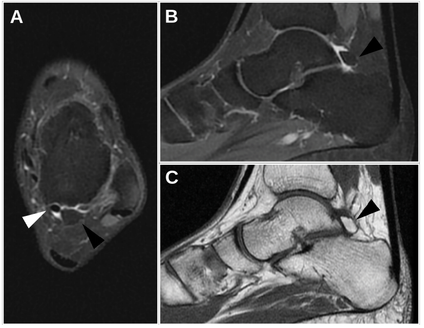
Images of an ankle MRI in fat-saturated proton density (PD) weighted (A, B) and T1-weighted (C) sequences, showing an accessory bone facing the posterior process of the talus, called “OS trigonum” (black arrow). The OS trigonum is prevalent in 2% to 50% of the population. When present, it sits just laterally to the groove of the flexor halluces longus (FHL) tendon. Thus, during plantar flexion, the OS trigonum with the FLH tendon can be impinged between the posterior surface of the tibia and the upper surface of the calcaneus, causing a LFH tenosynovitis, described as the OS trigonum syndrome.
Keywords: MRI; Trigonum; Tenosynovitis
 Awards Nomination