PHONE
+44-7482-878921
+44-7482-878921
2376-0249
Clinical-Medical Image - International Journal of Clinical & Medical Images (2024) Volume 11, Issue 1

Author(s): Nadia Boujida*, Soukaina Jabour, Omar El Aoufir, Fatima Zahra Laamrani, and Laila Jroundi
Department of Radiology, Ibn Sina University Hospital, Rabat, Morocco
*Corresponding Author:
Nadia Boujida
Department of Radiology
Ibn Sina University Hospital
Rabat, Morocco
Tel: +91 9912991421
E-mail: nadiaboujida21@gmail.com
Received: 08 November 2023, Manuscript No. ijcmi-24-119548; Editor assigned: 10 November 2023, Pre QC No. P-119548; Reviewed: 01 January 2024, QC No. Q-119548; Revised: 08 January 2024, Manuscript No. R-119548; Published: 15 January 2024, DOI:10.4172/2376-0249.1000931
Citation: Boujida N, Jabour S, Aoufir OEI, Laamrani FZ and Jroundi L. (2024) Thoracic Paravertebral Extramedullary Hematopoiesis. Int J Clin Med Imaging 10: 931.
Copyright: © 2024 Boujida N, et al. This is an open-access article distributed under the terms of the Creative Commons Attribution License, which permits unrestricted use, distribution, and reproduction in any medium, provided the original author and source are credited.
A 62-year-old adult with myelofibrosis presented with a chronic cough for six months, along with progressive chest pain and dyspnea, without any fever and in a preserved general condition. Clinical examination revealed pale conjunctivae and tachycardia.
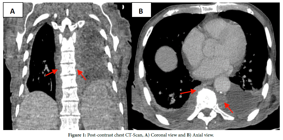
A post-contrast chest CT scan was performed, showing thickening of the bilateral thoracic para-vertebral soft tissues (red arrows), with bilateral pleural effusion and splenomegaly.
Extra Medullary Hematopoiesis (EMH) is defined as the abnormal formation of blood tissue outside the bone marrow. It is rarely primary, and although it can be secondary to bone or neoplastic pathologies, it is often secondary to a chronic hematological disorder with phenomena of medullary hyperstimulation. The four hematological disorders most commonly associated with EMH are idiopathic myelofibrosis, betathalassemia, chronic myeloid leukemia, and sickle cell disease. It is important to note that EMH primarily affects men and typically manifests around the ages of 60 to 70 [1].
This phenomenon occurs in various organs, including the liver, spleen, lymph nodes, kidneys, paravertebral regions, peritoneum, and pleural cavity [2].
In general, EMH does not present any clinical manifestations, except when it occurs in the epidural space, causing signs of spinal cord or radicular compression [3].
Para-vertebral EMH manifests as large masses of soft tissue in the thoracic paravertebral regions. These masses rarely cause significant symptoms, but they can lead to pleural effusion, hemothorax, or dyspnea.
The precise source of the hematopoietic tissue in this region remains uncertain. It has been suggested that hematopoietic tissue from the vertebral bone marrow may be extruded through weakened areas of trabecular bone into the epidural space, where it could proliferate.
On CT-scan, these lesions appear as paravertebral masses that do not erode the adjacent bone and have a density similar to that of the surrounding muscle. These lesions enhance uniformly after contrast injection due to their high vascularity. In cases of older lesions, iron deposition and fatty infiltration can lead to the appearance of a mass with inhomogeneous enhancement. MRI is a useful modality for evaluating these paravertebral masses. T1-weighted images may show areas of hyperintensity within the mass, which can be attributed to the presence of fat [4].
If clinical features or radiological signs of chronic anemia, such as rib enlargement and cortical thinning, are present, the diagnosis of EMH can be easily confirmed. In cases where there is no underlying hematological disorder or in uncertain cases, the diagnosis can be validated through a CT-guided biopsy or thoracoscopy [4].
Among the differential diagnoses, we can mention nerve tumors, mesothelioma, lipoma, pleural fibroma, and tuberculous abscesses [3].
Paravertebral EMH typically regresses after treatment with blood transfusions, hydroxyurea, or radiotherapy [4].
Extra-medullary hematopoiesis; EMH; Paravertebral EMH; Chest CT
None of the authors has any conflict of interest to disclose.
[1] Broucqsault, A., Ouzzane, A., Launay, D., Leroy, X. and Rose, C., et al. (2011). Hématopoïèse extramédullaire rénale. Adv Urol 21: 575-579.
Google Scholar, Crossref, Indexed at
[2] Rafiee, F., Haseli, S., Jafari, S. H., and Iranpour, P. (2020). Case report: Extramedullary haematopoiesis presenting as a periportal mass. BMJ Case Rep 13.
Google Scholar, Crossref, Indexed at
[3] A benhmouda. L’hématopoïèse extra-médullaire para-vertébrale. Research Journal. 2017-06-22.
[4] Sohawon, D., Lau, K. K., Lau, T. and Bowden, D. K. (2012). Extraâ?Âmedullary haematopoiesis: A pictorial review of its typical and atypical locations. J Med Imaging Radiat Oncol, 56: 538-544.
Google Scholar, Crossref, Indexed at
 Awards Nomination