PHONE
+44-7482-878921
+44-7482-878921
2376-0249
Clinical Image - International Journal of Clinical & Medical Images (2016) Volume 3, Issue 6

Author(s): Akira Baba, Yumi Okuyama, Shogo Kaida and Junichi Matsui
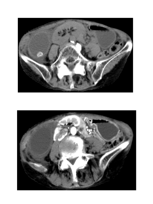
An otherwise-healthy 82-year-old woman presented to the emergency department with abdominal pain gradually migrating to the right and vomiting. Her body temperature was 37.2°C, abdominal examination showed tenderness in the right lower abdomen, and her WBC was 8,400/μL no plasma levels of enzymes were elevated. She was thin (body mass index 20.4 kg/m2 ) with scoliosis. Non-enhanced Computed Tomography (CT) revealed a massively swollen gallbladder with high dense (Figure 1) and contrast enhanced CT demonstrated poorly-enhanced edematous wall thickening (Figure 2), with twisted pedicle detached from the liver, suggesting gallbladder torsion. Emergent cholecystectomy was performed, revealing a 270° counterclockwise torsion of the gallbladder. Gallbladder torsion is more prevalent in thin elderly females with spinal deformation [1]. Characteristic contrast enhanced CT findings are poorly-enhanced and thickened gallbladder wall, pericholecystic fluid, and twisted pedicle (whirl sign) [2]. Contrast enhanced CT can help establish the preoperative diagnosis of gallbladder torsion in cases involving elderly females complaining abdominal pain.
 Awards Nomination