PHONE
+44-7482-878921
+44-7482-878921
2376-0249
Case Blog - International Journal of Clinical & Medical Images (2014) Volume 1, Issue 2

Author(s): Robert Evans Heithaus*
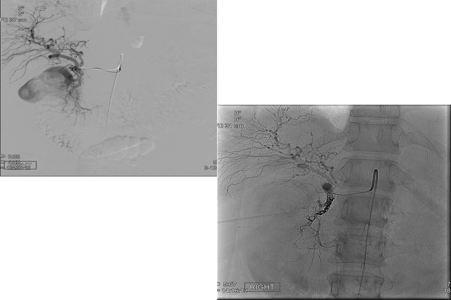
Upper Gastro Intestinal (UGI) bleeding is a common medical problem with significant associated morbidity and mortality. Prompt management is essential, and clinicians should familiarize themselves with the various treatment modalities including surgical and vascular interventional strategies. We present a case of acute UGI hemorrhage from duodenal ulcers treated by transarterial coil embolization in a patient who had previously undergone gastroduodenal arterial branch ligation. A 47 year-old female with known history of duodenal ulcers and prior surgical ligation of an arterial feeding branch from the gastroduodenal artery (GDA) presented to the emergency department with syncope. She was discovered to have massive upper gastrointestinal bleeding. The patient underwent intravenous fluid resuscitation including the transfusion of 4 units of packed red blood cells. Despite continued medical therapy the patient remained in hypovolemic shock. She was brought to the interventional suite for further management. The right femoral artery was punctured with a micropuncture technique; and a sheath was advanced into the artery over a wire. The celiac artery was selectively catheterized and digital subtracted angiography (DSA) was performed. Massive active arterial extravasation of contrast was observed from the gastroduodenal arterial distribution (Figure 1). A microcatheter was then advanced distally beyond the level of active arterial extravasation and coil embolization was begun (Figure 2). Multiple coils were deployed; however, post embolization angiography demonstrated persistent active arterial extravasation from an additional GDA branch (Figure 3). Selective micro catheterization of this arterial branch was undertaken and additional coils were deployed (Figure 4). Repeated post embolization arteriography was performed, and no active arterial extravasation was seen. The patient was had an uneventful postoperative course and was ultimately discharged from the hospital 7 days later in satisfactory condition.
Figure 1: Selective catheterization of the superior mesenteric artery was performed. Digital subtraction angiography demonstrates contrast extravasation from a gastroduodenal arterial (GDA) branch.
Figure 2: Multiple 0.018 platinum coils were deployed across the proximal and distal portion of the bleeding vessel.
Figure 3: Delayed DSA images demonstrate persistent contrast extravasation from an additional GDA branch.
Figure 4: Additional coils were placed, and follow up DSA revealed no additional areas of contrast extravasation.
Corresponding authr
Robert Evans Heithaus
Department of Radiology
Baylor University Medical Center
Dallas
USA
 Awards Nomination