PHONE
+44-7482-878921
+44-7482-878921
2376-0249
Research Article - International Journal of Clinical & Medical Images (2017) Volume 4, Issue 3

Author(s): Darwish HS1, Kameln HA and Habash MY
Abstract: To assess the usefulness of ultrasound in assessment and diagnosing foot peudotumoural soft tissue lesions.
Material and Methods: We conducted a retrospective study of a 43 patients who presented to the Dallah hospital Riyadh, Saudi Arabia, Radiology department, with foot swelling+pain. All our cases have X-ray foot as well as, muscol-sketetal ultrasound, results of MRI study, and biopsy results of some cases were included.
Results: The mean age of the patients was 16 ± 35 years, Females to male’s ratio was 4:1. The collected data of imaging US features, with exact lesion location, the relationship with surrounding structures and clinical features such as sex, age and symptoms in addition to X-ray, MRI findings as well as biopsy results if done allowing accurate diagnosis including; 9 cases (21%), Morton’s neuroma, (16%), 7 cases synovial cysts, 9 cases ganglion cysts (21%), 3 cases post traumatic cyst (7%), 3 cases marked subcutaneous edema (7%), 2 cases gouty arthritis with tophi formation (5%), 2 cases bursitis (5%), while tenosynovitis seen in 8 cases (18%).
Conclusion: Muscolo skeletal sonography is low in cost, easily available, highly efficient and can be used in the assessment of pseudo tumoural soft tissue lesions of the foot and ankle.
Keywords: Soft tissue; Pseudo tumours; Foot; Ultrasound; MRI; Musculoskeletal sonography
Introduction In the United States, approximately 40% of adults experience foot problems [1]. The foot can be affected by a variety of congenital, inflammatory, infectious, degenerative, and neoplastic disorders [2]. Foot soft tissue lesions include benign and malignant neoplasms, as well as non-neoplastic or pseudotumoural lesions [3]. The pseudotumoural lesions of the foot comprise a diverse group with widely varying histopathology; the most common non-tumoral lesions are benign cysts, posttraumatic, reactive, and inflammatory lesions. These lesions include synovial cysts, ganglion cysts, tenosynovitis, epidermoid cysts, masses (abscess, necrosis and hematoma) that show cystic transformation, gout tophi, rheumatoid nodules, Morton’s neuroma and granuloma annulare [3.4]. Aetio-pathogenesis of these lesions is varied, but in general degenerative, inflammatory and infectious phenomena may play a role [3]. Ganglion and synovial cysts are the most common soft tissue lesions in the ankle and foot region, most frequently located around the ankle or at the dorsum of the foot [5]. Morton’s neuroma (fibroma) is a common cause of intermetatarsalgia, ultrasound examination is reliable in detecting Morton’s fibroma [6]. MR imaging has been proven to be highly sensitive and specific in diagnosing Morton’s fibroma, as well as providing an accurate preoperative assessment [7]. Tenosynovitis involves inflammation of the tendon sheath. The etiology includes acute and chronic trauma, infection or inflammatory or metabolic disease. Excessive fluid accumulation in the tendon sheath leads to a mass-like presentation [8]. Soft tissue lesions of the foot and ankle are a relatively rare cause of referral for medical imaging [3]. MR imaging is being recognized as the modality of choice for assessment of pathologic conditions of the ankle and foot, it demonstrates abnormalities in the bones and soft tissues before they become evident at other imaging modalities [9]. However, because of advances in technology, sonography offers significant advantages over other imaging techniques in assessing muscle trauma. High-frequency transducers yield images with excellent spatial resolution. The real-time capability allows dynamic evaluation of muscle and tendon injuries [4].
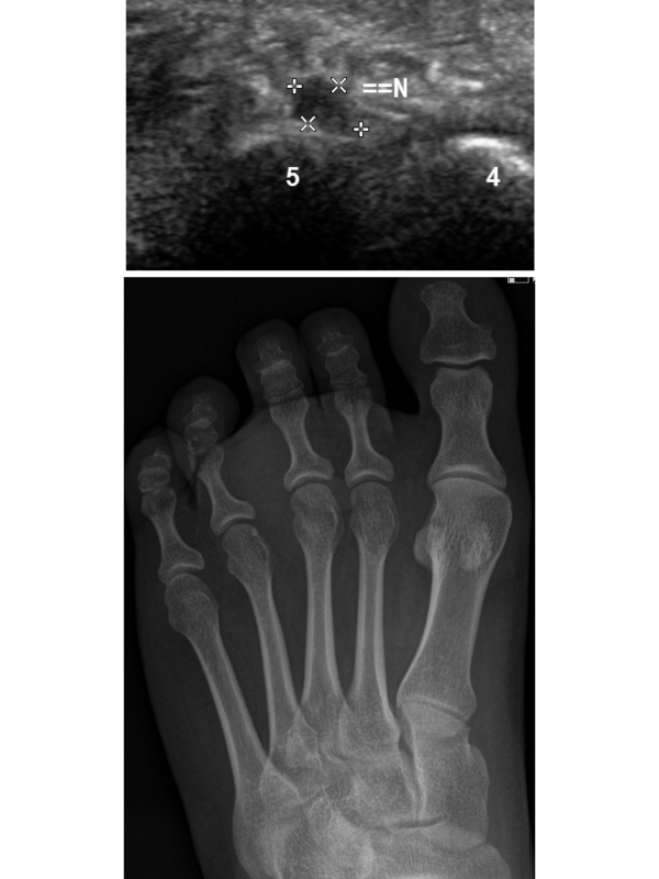
Results: In our study, 43 patients were included, foot swelling was main complain, associated pain was seen in 38 patients (88%). The mean age of the patients was 16 ± 35 years, Females to males’ ratio was 4:1. Associated complaints in included cases were pain in other regions of the leg such as the knee and thigh (21%), soft masses (14%), and leg swelling (2%). The duration of symptoms until presentation was less than one month in 29 cases (67%) and more than 1 month in 14 cases (33%). Morton’s neuroma seen in 9 cases (21%), all cases confirmed diagnosis with biopsy and MRI included for all 8 cases. All our 9 patients are middle age females in 2 of them the lesion was less than 5 mm diameter (Figure 1), while in 7 cases, the lesion is more than 5 mm (Figure 2) and all are complaining of web space pain radiating to the toe and the pain is worsen by wearing shoes and walking. 6 cases found in third inter metatarsal space while the other 2 cases found in 2 inter metatarsal space and only one case noted in 4th web space. In our study 7 cases (16%) diagnosed as synovial cysts (Figure 3), and 9 cases diagnosed as ganglion cysts (21%) (Figure 4), most of cases were simple cysts; however, 1 of 7 cases and 2 of 9 cysts were complicated and demonstrate partially or completely hyper echogenic content (Figure 5). In our study 3 cases showing marked subcutaneous edema (7%) leading to foot swelling more marked at medial aspect (Figure 6). Sonography revealed 3 cases (7%) post traumatic cystic lesions (Figure 7), one of them less than 1 week duration and the other 2 cases more than one week duration. Sonography also revealed tenosynovitis (Figure 8) in 8 cases (18%), tibialis anterior tendon is the most affected tendon 5 cases, 2 cases showing tibialis posterior tenosynovitis. Peroneal tenosynovitis was seen in 1 patient. Gouty arthritis was found in 2 cases (5%) with tophi formation, another 2 cases (5%) showing sonographic features of bursitis (Figure 9).
 Awards Nomination