PHONE
+44-7482-878921
+44-7482-878921
2376-0249
Clinical-Medical Image - International Journal of Clinical & Medical Images (2023) Volume 10, Issue 2

Author(s): Dabiah Alboaneen*
Department of Urology, Ultragen Medical Center, 31-572 Krakow, Poland
Received: 19 January 2023, Manuscript No. ijcmi-23-96525; Editor assigned: 20 January 2023, Pre QC No. P-96525; Reviewed: 09 February 2023, QC No. Q-96525; Revised: 14 February 2023, Manuscript No. R-96525; Published: 21 February 2023, DOI:10.4172/2376-0249.1000880
Citation: Alboaneen D. (2023) Understanding Clinical Medical Image Analysis in Modern Healthcare: An Overview. Int J Clin Med Imaging 10:880.
Copyright: © 2023 Alboaneen D. This is an open-access article distributed under the terms of the Creative Commons Attribution License, which permits unrestricted use, distribution, and reproduction in any medium, provided the original author and source are credited.
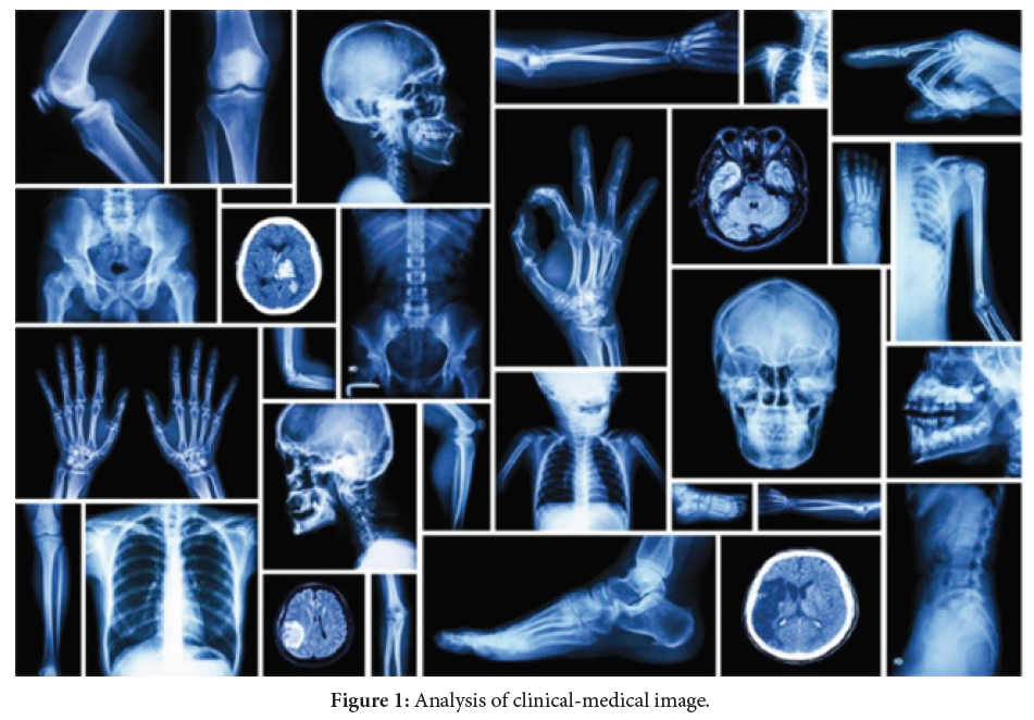
Clinical medical image analysis refers to the process of extracting meaningful information from medical images, such as X-rays, CT scans, MRI scans and ultrasound images. The goal of clinical medical image analysis is to aid in the diagnosis, treatment and monitoring of medical conditions by providing quantitative measurements and visual representations of anatomical structures and physiological functions. Medical image analysis involves a range of techniques and methods, including image preprocessing, segmentation, registration, feature extraction and classification. Preprocessing involves techniques such as noise reduction, intensity normalization and image enhancement, to improve the quality of the images and facilitate subsequent analysis. Segmentation refers to the process of separating an image into meaningful regions, such as organs or tissues, while registration involves aligning multiple images taken at different times or from different imaging modalities. Feature extraction involves identifying and quantifying characteristics of the image, such as texture, shape and intensity, which can be used to distinguish between different tissues or identify specific anatomical structures. Classification involves using machine learning algorithms to assign a label or diagnosis based on the features extracted from the image. Clinical medical image analysis has a wide range of applications, including the detection and diagnosis of diseases such as cancer, cardiovascular disease and neurological disorders. It can also be used for treatment planning, monitoring disease progression and evaluating the effectiveness of interventions. Overall, clinical medical image analysis plays a critical role in modern healthcare by providing clinicians with quantitative measurements and visual representations of medical conditions, allowing for earlier and more accurate diagnoses and more effective treatments [1,2].
Artificial intelligence; Colorectal cancer; Deep learning; Early diagnosis; Machine learning
None of the authors has any conflicts of interests to disclose.
[1] Wang XL, Huang HY, Li Z, Yu YS and Hu YQ, et al. (2015). Risk factors associated with aortic remodeling in patients with Stanford type B aortic dissection after thoracic endovascular aortic repair. Genet Mol Res 14: 11692-11699.
Google Scholar, Crossref, Indexed at
[2] Wojciechowski J, Znaniecki L, Kaszubowski M and Rogowski J. (2019). Late aortic remodeling after endovascular repair of complicated type B aortic dissection—TEVAR protects only the covered segment of thoracic aorta. Ann Vasc Surg 55: 148-156.
 Awards Nomination