PHONE
+44-7482-878921
+44-7482-878921
2376-0249
Clinical-Medical Image - International Journal of Clinical & Medical Images (2021) Volume 8, Issue 5

Author(s): Armel Junior Tokpo, Baderddine Mohammadine, Kaoutar Stitou, Faycal Lakhdar, Mohammed Benzagmout, Khalid Chakour, Mohammed El-Faiz Chaoui
Clinical Image
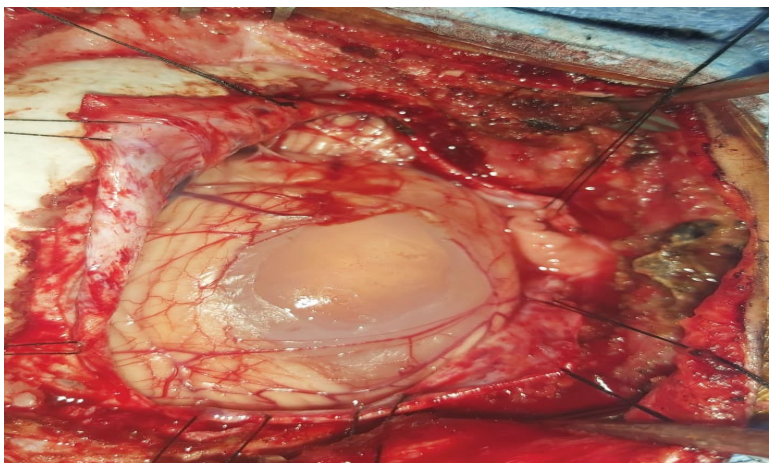
We report the case of a 5-years-old boy, presenting progressive headaches and mild cerebellar syndrome (gait disturbance, dysmetria) without significant burden on life’s quality. Funduscopic examination revealed grade 2 papilledema. Brain CT scan disclosed a left cerebellar hypodense unenhanced formation compatible with hydatid cyst (Figure 1). No others location had been found on chest, abdominal and pelvic computed tomography scan. During surgery, the cyst came to view after sub occipital craniotomy and dura opening (Figure 2) disrupting the cortical surface of the left cerebellum hemisphere. Complete delivering is achieved without rupture (Figure 3). Hydatid disease is a worldwide disease caused by Echinococcus tapeworm. Two species (Echinococcus granulosis and Echinococcus multilocularis) are linked with humans [1]. Among possible locations, intracranial hydatid cysts are rare (1% to 2%) and posterior fossa’s ones are exceptional [1,2]. This affection is an endemic disease in Morocco and children between 5-8 years old are much more concerned [2]. Multiple symptoms related to posterior fossa hydatid cyst could be encountered. Cerebellar syndrome, delayed intracranial hypertension signs (headaches, vomiting, seizures…), cranial nerve deficits [2] or long pathways signs. Brain CT scan is a gold standard for approaching the diagnosis. Typically (as in this case) it is hypodense well limited intra parenchymatous formation without peripheral edema and enhancement after gadolinium injection [2]. Surgical evacuation of the cyst is mandatory and includes different options (hydraulic dissection of Arana Iniguez, ponction-aspiration of the cyst). In this case the patient was secured to the operating table. Gradual table tilting associated with hydraulic dissection and gentle manual pression to roll the cyst out of the cerebellum have been conducted with success. Post operatively the boy was doing well and medical treatment with Albendazole has been conducted.
Keywords: Posterior fossa; Hydatid cyst; Child
 Awards Nomination