PHONE
+44-7482-878921
+44-7482-878921
2376-0249
Clinical-Medical Image - International Journal of Clinical & Medical Images (2022) Volume 9, Issue 4

Author(s): Benayad Aourarh*, Khadija Boualiten and Mouna Tamzaourte
Received: 25 March, 2022, Manuscript No. ijcmi-22-62359; Editor assigned: 27 March, 2022, 2022, PreQC No. P-62359; Reviewed: 14 April, 2022, QC No. Q-62359; Revised: 19 April, 2022, Manuscript No. R-62359; Published: 26 April, 2022, DOI: 10.4172/2376-0249.1000820
Clinical-Medical Image
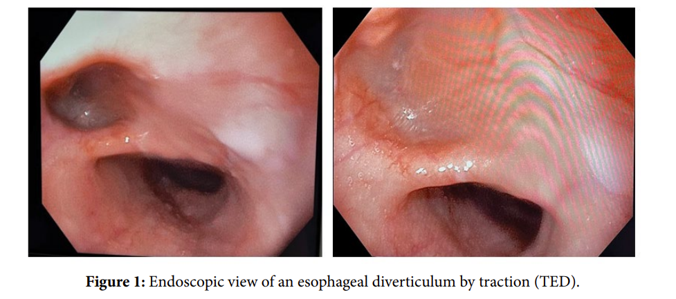
A seventy-nine-year-old healthy woman, with a history of hypertension, was referred to our institution for dysphagia exploration. She reported a one-year history of progressive dysphagia to solids with a recent episode of solid food getting stuck in her throat. She was a not a smoker, nor an alcohol user. She had no known history of esophageal dysmotility disorder and no history of diabetes or digestive cancer in her family. An upper gastrointestinal endoscopy was performed revealing a large esophageal diverticulum 28 cm from the incisors (Figure 1), without any ulceration. The rest of the endoscopy was otherwise normal. The diagnosis of a traction esophageal diverticulum (TED) was retained. Esophageal diverticulum is a relatively rare disorder of the esophagus [1], with prevalence up to 3% thanks to radiological and endoscopic findings [2]. Traction esophageal diverticulum can be defined as a pouch that protrudes externally, in a weak portion of the esophageal lining [3]. They usually appear in the middle one third of the thoracic esophagus and are attributed to radial traction from mediastinal inflammatory processes (such as tuberculosis or histoplasmosis). This reaction results in a paraoesophageal thickness pinching on the esophageal wall, leading to a localized diverticulum [4]. Usually asymptomatic, 15 to 20% of the patients can develop symptoms such as dysphagia, postural regurgitations, epigastric pain and heartburn. Most patients are diagnosed by barium esophagogram or an upper gastrointestinal endoscopy. A manometry can also be performed as it helps to rule out motility disorders. Management of TED include treating the underlying cause and local excision of the diverticulum (via thoracotomy or thoracoscopy) in symptomatic patients [5].
Keywords: Dysphagia; Esophagus; Diverticulum
References
[1] Hussain T, Maurer JT, Lang S, & Stuck B A (2017) Pathophysiologie, Diagnose und Therapie des Zenker-Divertikels. HNO 65:167-176.
Google Scholar Crossref Indexed at
[2] Hoghooghi D, Coakley FV, Breiman RS, Qayyum A, & Yeh BM (2006) Frequency and etiology of midesophageal diverticula at barium esophagography. Clin Imaging 30:245-247.
Google Scholar Crossref Indexed at
[3] Yam J, Baldwin D, & Ahmad SA (2021) Esophageal Diverticula. n: StatPearls. StatPearls Publishing, Treasure Island (FL)
[4] Ballehaninna UK, Shaw JP, & Brichkov I (2012) Traction esophageal diverticulum: A rare cause of gastro-intestinal bleeding. Springerplus, 1:1-2.
Google Scholar Crossref Indexed at
[5] Mitchell KG, Corsini EM, Van Haren RM., Walsh GL, & Sepesi B (2019) A case report of a midesophageal diverticulum mimicking a fibrovascular esophageal polyp. Int J Surg Case Rep 59:205-207.72.
Google Scholar Crossref Indexed at
 Awards Nomination