PHONE
+44-7482-878921
+44-7482-878921
2376-0249
Clinical-Medical Image - International Journal of Clinical & Medical Images (2022) Volume 9, Issue 3

Author(s): El Houss Salma*, Zebbakh Hajar , Lrhorfi Najlae , EL Haddad Siham , Chat Latifa, Allali Nazik
Received: 21 March 2022, Manuscript No. ijcmi-22-60201; Editor assigned: 23 March 2022, 2022, PreQC No. P-60201; Reviewed: 25 March 2022, QC No. Q-60201; Revised: 27 March 2022, Manuscript No. R-60201; Published: 30 March 2022, DOI: 10.4172/2376-0249.1000819
Clinical-Medical Image
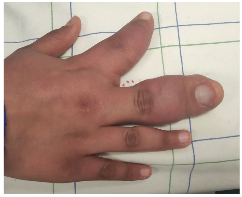
Marcrodystrophia lipomatosa is a rare congenital anomaly characterized by an abnormal growth of the mesenchymal tissues of one or more digits (usually the 2nd or 3rd digit) of the same limb (hand or foot); the damage can sometimes be bilateral or involve a whole limb [1] This gigantism is secondary to an abnormal proliferation of the adipose tissue of the mesenchymal elements involving the subcutaneous tissue, the muscle, the nerve sheath, the bone marrow and the periosteum. It is a non-hereditary pathology whose physiopathology is still poorly elucidated, The sex ratio of this disease is one, two subtypes have been described: the static type where the growth of the hypertrophied finger progresses at the same rate as that of the other fingers and the progressive type where the growth of the hypertrophied finger progresses more rapidly than that of the other fingers [2]. Standard radiography (Figure 1) can detect soft tissue and bone hypertrophy, the presence of radiolucent areas due to the presence of adipose tissue. On the CT scan (Figure 2), the excessive growth of bone and the proliferation of adipose tissue in the muscle fibers of the area concerned can be used as a guide to the diagnosis. Magnetic resonance imaging (Figure 3) is pathohgnomonic and can easily demonstrate fatty infiltration (T1 hypersignal with fat saturation on DP-STIR sequence) of subcutaneous tissue, muscle and nerve branches, Linear hypointense fibrous bands can be noted in this abnormal fat corresponding to the nerve bundles, bone hypertrophy and cortical thickening in the affected body part may lead to exostoses as bony outgrowths of the involved bone [1]. In addition to the interest of MRI in the positive diagnosis (Figure 4), it also allows the exclusion of other differential diagnoses such as: Neurofibromatosis type I, lymphangiomatosis, Klippel-Trenaunay disease [3], Hemangiomatosis and Proteus syndrome. The treatment is surgical and is performed for aesthetic purposes while preserving neurological function.
Keywords: Neonatal macrodactyly; Bone hypertrophy; MRI
[1] Khan RA, Wahab S, Ahmad I, & Chana RS (2010). Macrodystrophia lipomatosa: Four case reports. Ital J Pediatr 36: 1-5
Google Scholar Crossref Indexed at
[2] Rosean SCB, & Castleman B. (1958) LIEBOW AA: Pulmonary alveolar proteinosis. New Eng J Med 258: 1123-1125.
Google Scholar Crossref Indexed at
[3] Prabhu CS, Madhavi K, Amogh VN (2019). Macrodystrophia lipomatosa: A single large radiological study of a rare entity. J Clin Imaging Sci 9: 55-59.
 Awards Nomination