PHONE
+44-7482-878921
+44-7482-878921
2376-0249
Clinical Image - International Journal of Clinical & Medical Images (2014) Volume 1, Issue 9

Author(s): Shivali V Kashikar*
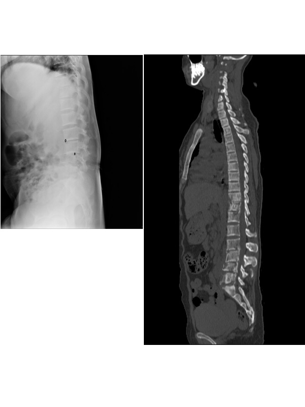
A postnatal 24 year old postnatal female presented to the emergency department with low grade fever, fatigue and backpain. Investigations revealed Sickle cell disease with haemoglobin of 5 g/dL and decreased hematocrit (29%). Haemoglobin electrophoresis showed Hb F- 38.3%, Hb S- 61.2% and Hb A2- 0.5%. White cell count was elevated and peripheral blood smear
demonstrated target cells but no sickle shaped erythrocytes. Radiograph of the dorso-lumbar spine (Figure 1) showed diffuse ill defined mixed lytic-sclerotic lesions in vertebral bodies and destruction around the end plates. Computed tomography (Figure 2) confirmed the same. Magnetic Resonance Imaging (MRI) demonstrated extensive but patchy and geographic pattern of signal abnormalities in the dorsal and lumbar vertebrae, heterogeneous on T1 and T2 weighted imaging. The lesions in L3 and L4 vertebrae showed an enhancing rim (Figure 3). The diagnosis of bone marrow necrosis (BMN) was confirmed by trephine biopsy of L4 vertebra which showed extensive destruction of haematopoietic tissue and the stroma with preserved bone. She was treated conservatively with blood transfusion and analgesia. BMN in a patient with sickle cell disease (SCD) was first described by Wade and Stevenson, although it represents only 2% of all BMN reported cases [1]. Extensive bone marrow necrosis is a
rare but severe complication of sickle cell disease and is also an unusual presenting feature in a young adult [2]. The preferred modality to evaluate diseases of the bone marrow is Magnetic Resonance imaging (MRI) [3]. It is a non-invasive technique that complements bone marrow aspirations and biopsies by providing the signal characteristics of trabecular bone, fat, and water.
MRI demonstrates the characteristic diffuse, extensive and geographic pattern of signal abnormalities in the vertebrae. It consists of a central area of variable signal intensity that is surrounded by a distinct peripheral enhancing rim. BMN has been seen in hematologic malignancy, sickle cell disease, infection, and adverse effects of medications [4].
Generalized BMN has also been observed in patients with sickle cell disease (SCD), where it is due to blood vessel occlusion and its diagnosis requires a bone marrow biopsy. Systemic fat embolism and acute multi-organ failure syndrome can further complicate the clinical scenario of BMN in patients with SCD. The prognosis of generalized BMN is slightly better in SCD and
other non-malignant states as compared to that seen in leukaemia [5]. Bone marrow necrosis (BMN), avascular necrosis (AVN) and marrow aplasia of bone are distinct entities. There is disruption of the normal marrow architecture and necrosis of myeloid tissue in BMN. It is an extensive process and affects the marrow of the spine and pelvis [6]. The lesions of BMN do not usually progress to collapse of vertebral body. On the other hand, AVN and bone infarcts are focal and commonly found in periarticular locations and long bones, especially the femoral head, but rarely in the vertebra [7]. Vertebral body collapse in seen in later stages of AVN.
 Awards Nomination