PHONE
+44-7482-878921
+44-7482-878921
2376-0249
Clinical-Medical Image - International Journal of Clinical & Medical Images (2021) Volume 8, Issue 8

Author(s): Jihane El-Mandour*, Laila Jroundi, and Laamrani FZ
Clinical Image
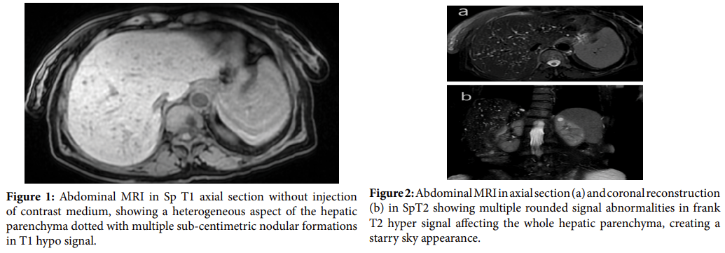
Von Meyenburg complexes (VMCs), also known as biliary micro hamartomas, were first described in 1918, and are generally considered to be a benign condition due to a developmental abnormality of the ductal plate. This hypothesis is supported by the fact that Von Meyenburg complexes (VMCs) are frequently observed in association with other conditions related to ductal plate malformation, such as Caroli disease and congenital hepatic fibrosis. Von Meyenburg complexes (VMCs) are usually diagnosed incidentally by imaging through the individualization of multiple small cystic lesions. They usually measure between 1 and 15 mm in diameter, rarely they can exceed 10 cm, becoming symptomatic. When they are small, they may escape imaging. The sonographic appearance of Von Meyenburg complexes (VMCs) is variable and non-specific. The hepatic parenchyma is heterogeneous. VMCs present as multiple micro nodules, hypo- or hyperechoic, these micronodules are often very small and may present comet tail artifacts, which makes them difficult to differentiate from aerobilia and intrahepatic lithiasis. On CT scan, VMCs appear as multiple round or irregular hypodense lesions that do not enhance after injection of contrast medium. They may be scattered in the liver parenchyma, but mainly in the subscapular and periportal areas. However, they remain difficult to characterize because of their small size, infra centimetric. On MRI, VMCs are hypo signal on T1-weighted sequences (Figure 1), and frankly hyper signal on T2-weighted sequences (Figure 2). [VMCs are often irregular in shape with well-defined margins. On the diffusion sequence, they behave similarly to cystic lesions. Cholangio-MRI in VMCs does not show communication of the cystic formations with the bile ducts contrary to Caroli disease where this communication exists. MRI and Cholangio-MRI are of ultimate help in the diagnosis of CMVs and differentiate them from other entities. The differential diagnosis is mainly with cystic forms of liver metastases, polycystic liver disease, Caroli disease; biliary cysts, congenital liver fibrosis, and autosomal dominant polycystic liver disease. There are rare cases of associated cholangiocarcinoma; however, no routine follow-up imaging is recommended. The presence of VMCs may prompt careful consideration of the coexistence of cholangiocarcinoma and the search for cystic kidney disease.
 Awards Nomination