PHONE
+44-7482-878921
+44-7482-878921
2376-0249
Clinical-Medical Image - International Journal of Clinical & Medical Images (2023) Volume 10, Issue 4

Author(s): Halfi Mohamed Ismail*, Imrani Kaoutar, Nabil Moatassim Billah and Nassar Ittimade
Department of Central Radiology, Ibn Sina University Hospital Center, Rabat 10000, Morocco
Received: 12 March 2023, Manuscript No. ijcmi-23-92476; Editor assigned: 13 March 2023, Pre QC No. P-92476; Reviewed: 29 March 2023, QC No. Q-92476; Revised: 05 April 2023, Manuscript No. R-92476; Published: 12 April 2023, DOI:10.4172/2376-0249.1000887
Citation: Ismail HM, Kaoutar I, Billah NM and Ittimade N. (2023) Xanthogranulomatous Cholecystitis: Role of High Resolution Ultrasound (HRUS) in the Diagnosis. Int J Clin Med Imaging 10:887.
Copyright: © 2023 Ismail HM, et al. This is an open-access article distributed under the terms of the Creative Commons Attribution License, which permits unrestricted use, distribution, and reproduction in any medium, provided the original author and source are credited.
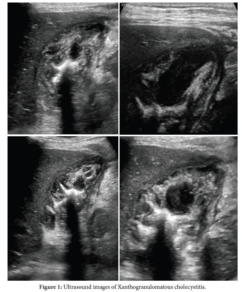
Mr. MB is a 51-year-old male presenting upper right quadrant pain with mild sign of jaundice, fever, and a clinical picture suggestive of gallbladder disease. A blood panel revealed an elevation of white blood cell count (WBC) at 20,000 μL. C-reactive protein was found to be 60 mg/L. Liver function tests (LFTs) showed elevated alkaline phosphatase (ALP), gamma-glutamyl transferase (GGT) and total bilirubin, each respectively at: 152 U/L, 71 U/L and 1.8 mg/dl. An abdominal ultrasound was performed which showed that the margin between the gallbladder lumen and mucosa was underlined with hyperechoic foci with and without comet-tails. Additionally, there were irregular hypoechoic intramural nodules distributed in a parallel fashion inside the wall of the gallbladder separated by hyper-echoic lines also known in literature as the ‘palisade presentation; a radiological hallmark of xanthogranulomatous cholecystitis. Multiple macro gallstones along with a 10 mm thick gallbladder wall were also observed among the lesions (the largest measuring 20 mm by 05 mm). A biopsy was performed to further confirm the diagnosis and which came back positive [1,2].
Xanthogranulomatous cholecystitis (XGC) is a scarce subtype of cholecystitis, distinguished by the accumulations of xanthogranulomas within the gallbladder wall thus responsible for its thickening and the destruction of its normal architecture [3]. It can lead to symptoms such as bilary colics, fever and jaundice. Due to the rarity of XGC and its ability to imitate other inflammatory infections of the gallbladder, it can be extremely difficult to diagnose. The diagnosis of XGC is usually confirmed through pathological findings. High resolution ultrasound (HRUS) has been the catalyst for detailed sonography images reflecting the micro-architecture of the soft tissues and structures within the body. The predictive accuracy of HRUS has been challenged in differentiating between gallbladder cancer and other inflammatory affluences of the gallbladder. First Kim and al in 2013 concluded that some ultrasonic characteristics of XGC can facilitate the differential diagnosis between XGC and GBC including smooth mucosa, small hypo-echoic nodules, hyper-echoic foci vs. an irregular mucosa, no inner wall nodules found in early stages of GBC before any malignant expansion at expense of the liver parenchyma [1,2]. Secondly Fan Zhang and al. in a 2019 found that based on all those features, now depicted thanks to HRUS, the positive predictive value in diagnosing XGC is around 75% with a 100% negative predictive value in radiologically setting apart XGC and GBC [1] (Figure 1).
The introduction of HRUS is a valuable tool in the diagnosis of XGC, and that by providing images with higher resolution and greater detail than traditional ultrasound. Therefore HRUS helps avoiding inappropriate management of the condition by decreasing the rate of unneeded cholecystectomies performed in patient with a false GBC diagnostic tag. Lastly, multiple AI models based on CT and MRI radiological features and diagnostic prediction of XGC showed good accuracy for the preoperative discrimination of XGC and GBC [4].
Xanthogranulomatous cholecystitis; Ultrasound; HRUS
None of the authors has any conflicts of interests to disclose.
[1] Zhang F, Chen W, Zhang L, Hou C and Zhang M. (2019). Usefulness of ultrasound in differentiating xanthogranulomatous cholecystitis from gallbladder carcinoma. Ultrasound Med Biol 45: 2925-2931.
Google Scholar, Crossref, Indexed at
[2] Kim JH, Lee JY, Baek JH, Eun HW and Kim YJ, et al. (2015). High-resolution sonography for distinguishing neoplastic gallbladder polyps and staging gallbladder cancer. Am J Roentgenol 204: W150-W159.
Google Scholar, Crossref, Indexed at
[3] Giudicelli X, Rode A, Bancel B, Nguyen AT and Mabrut JY. (2021). Xanthogranulomatous cholecystitis: Diagnosis and management. J Visc Surg 158: 326-336.
Google Scholar, Crossref, Indexed at
[4] Zhou QM, Liu CX, Zhou JP, Yu JN and Wang Y, et al. (2022). Machine learning-based radiological features and diagnostic predictive model of xanthogranulomatous cholecystitis. Front Oncol 12.
 Awards Nomination