PHONE
+44-7482-878921
+44-7482-878921
2376-0249
Clinical Image - International Journal of Clinical & Medical Images (2016) Volume 3, Issue 7

Author(s): Teruo Iwasaki, Son Chihyon and Yoshio Yamasaki
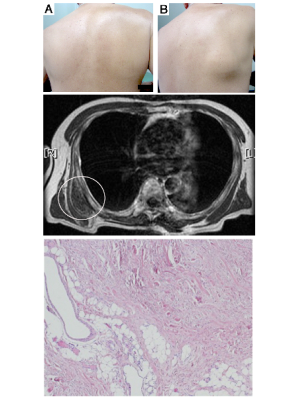
A 65-year-old man presented with a right subscapular mass with discomfort, snapping of the scapula and restriction of shoulder motion on the right side. His hobby was playing golf. He underwent left lobectomy for goiter at 34 years of age. On physical examination, with shoulder and arm in the neutral position, a projecting finding inferior scapula was normal (Figure A). On horizontal adduction of the right shoulder, a fist-sized mass appeared in the subscapular region (Figure B). Laboratory findings were unremarkable. Computed tomography revealed an approximately 8 cm heterogeneous soft-tissue mass in the infrascapular region. Magnetic resonance imaging showed a mass, which was isointense to skeletal muscle and contained funiform signals similar to adipose tissue (Figure C). The tumor was diagnosed as elastofibroma dorsi. Complete excision was indicated due to his symptom and the tumor size. Microscopically, the tumor was composed of eosinophilic dense collagen fiber bundles and elastic fibers, consistent with elastofibroma dorsi (Figure D). Postoperative course was uneventful, and no recurrence was found at 30 months after surgery. Elastofibroma dorsi is an uncommon benign soft tissue tumor with a characteristic location and imaging appearance [1, 2]. Professionals of clinical and medical fields should consider this condition in the differential diagnosis of a subscapular mass.
 Awards Nomination