PHONE
+44-7482-878921
+44-7482-878921
2376-0249
Clinical-Medical Image - International Journal of Clinical & Medical Images (2023) Volume 10, Issue 2

Author(s): Roberto Moretto*
Department of Surgery, Azienda Ospedaliero-Universitaria Pisana, Via Paradisa 2, 56124 Pisa, Italy
Received: 20 January 2023, Manuscript No. ijcmi-23-96524; Editor assigned: 21 January 2023, Pre QC No. P-96524; Reviewed: 13 February 2023, QC No. Q-96524; Revised: 18 February 2023, Manuscript No. R-96524; Published: 27 February 2023, DOI:10.4172/2376-0249.1000879
Citation: Moretto R. (2023) Imaging of Liver Transplants Procedure by MRI. Int J Clin Med Imaging 10:879.
Copyright: © 2023 Moretto R. This is an open-access article distributed under the terms of the Creative Commons Attribution License, which permits unrestricted use, distribution, and reproduction in any medium, provided the original author and source are credited.
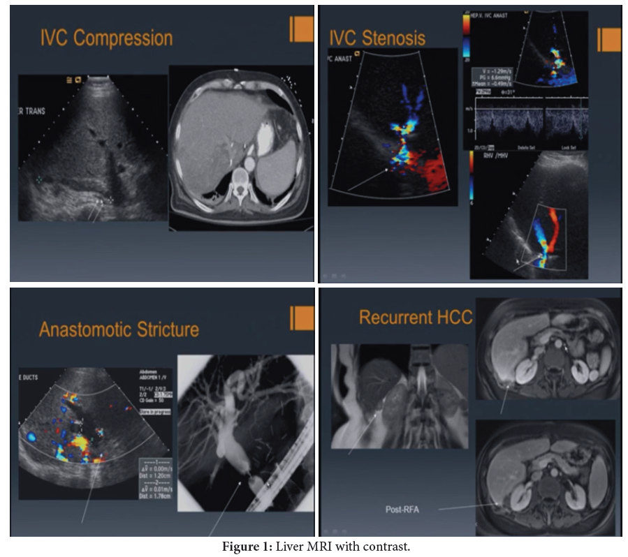
Liver transplantation is a life-saving surgical procedure for patients with end-stage liver disease or liver failure. Post-transplant monitoring is critical to assess graft function and identify potential complications. Imaging plays a vital role in the evaluation of liver transplant recipients. Multiple imaging modalities, such as ultrasound, computed tomography (CT), magnetic resonance imaging (MRI) and positron emission tomography (PET), can be used for imaging liver transplants. Ultrasound is the most common modality used for initial evaluation and follow-up imaging due to its noninvasiveness, cost-effectiveness and ability to provide real-time images. CT and MRI are more sensitive than ultrasound for detecting post-transplant complications, such as vascular or biliary complications and can provide high-resolution images of the transplanted liver. PET imaging can provide functional information and help distinguish between post-transplant complications and normal postoperative changes. Imaging findings must be correlated with clinical data to ensure accurate diagnosis and management of post-transplant complications. Imaging can also be used for pre-transplant evaluation of potential donors and recipients, as well as for planning surgical approaches and identifying anatomical variations [1,2].
Liver transplantation; Cholestasis; X-linked primary immunodeficiency
None of the authors has any conflicts of interests to disclose.
[1] Cohen P, Cross D and Jänne PA. (2021). Kinase drug discovery 20 years after imatinib: progress and future directions. Nat Rev Drug Discov 20: 551-569.
Google Scholar, Crossref, Indexed at
[2] Laufer S, Bajorath J, Gehringer M, Gray N and Frye S, et al. (2022). Publication Criteria and Requirements for Studies on Protein Kinase Inhibitors─ What Is Expected? (“It is pretty easy to make a bad kinase inhibitor”). J Med Chem 65: 6973-6974.
 Awards Nomination