PHONE
+44-7482-878921
+44-7482-878921
2376-0249
Clinical-Medical Image - International Journal of Clinical & Medical Images (2022) Volume 9, Issue 6

Author(s): Jihad Boularab*, Hind Sahli, Zineb Izi and Meriem Edderai
Department of Radiology, Mohammed V Military Hospital, Rabat, Morocco
Received: 06 June 2022, Manuscript No. ijcmi-22-66818; Editor assigned: 08 June 2022, Pre QC No. P-66818; Reviewed: 17 June 2022, QC No. Q-66818; Revised: 21 June 2022, Manuscript No. R-66818; Published: 28 June 2022, DOI: 10.4172/2376-0249.1000833
Citation: Boularab J, Sahli H, Izi Z and Edderai M. (2022) Pelligrini Stieda Syndrome: A Rare Complication of Knee Entorse. Int J Clin Med Imaging 9:833.
Copyright: © 2022 Boularab J, et al. This is an open-access article distributed under the terms of the Creative Commons Attribution License, which permits unrestricted use, distribution, and reproduction in any medium, provided the original author and source are credited.
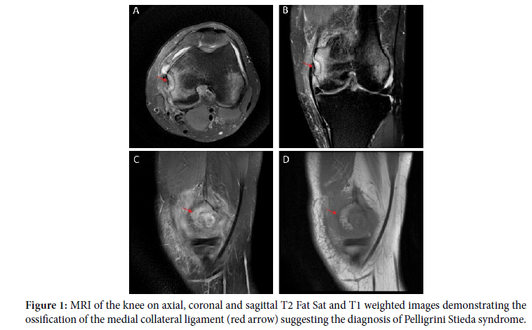
Axial, coronal and sagittal views of knee MRI of a 56 years old man with recurrent knee pain following a knee sprain, showing a calcification in the soft tissue situated mesial to the medial femoral condyle (red arrows (Figure 1)) corresponding to the ossification of the medial collateral ligament, suggesting the diagnosis of Pelligrini Stieda syndrome. The Pellegrini-Stieda syndrome is relatively infrequent and is commonly associated with sporting injuries. It occurs within 11 days to 6 weeks after trauma and patients may be asymptomatic or present a local pain and swelling in the medial aspect of the knee.
The diagnosis can be suggested on the X-ray, and confirmed by the MRI which also delineates the extent of adhesion of the calcified mass to the MCL and the remaining ligamentous volume [1,2] (Figure 1).
Pelligrini stieda; Posttraumatic; MRI; Ossification; Medial collateral Ligament
Google Scholar, Crossref, Indexed at
 Awards Nomination